← cell membrane coloring Cell membrane coloring (2) cell membrane bilayer The action potential · anatomy and physiology →
If you are looking for Transmission Electron Micrograph Of An Animal Cell Photograph by Dr you've came to the right place. We have 35 Images about Transmission Electron Micrograph Of An Animal Cell Photograph by Dr like The Cell: The Histology Guide, Membrane Architecture | Celebrate Cytochemistry | Gwen V. Childs, Ph.D. and also Cell Membrane Micrograph Stock Photos & Cell Membrane Micrograph Stock. Read more:
Transmission Electron Micrograph Of An Animal Cell Photograph By Dr
 pixels.com
cell electron micrograph animal transmission gopal murti dr photograph nuclear envelope 2nd uploaded which may science
Cell Membrane Micrograph High Resolution Stock Photography And Images
 www.alamy.com
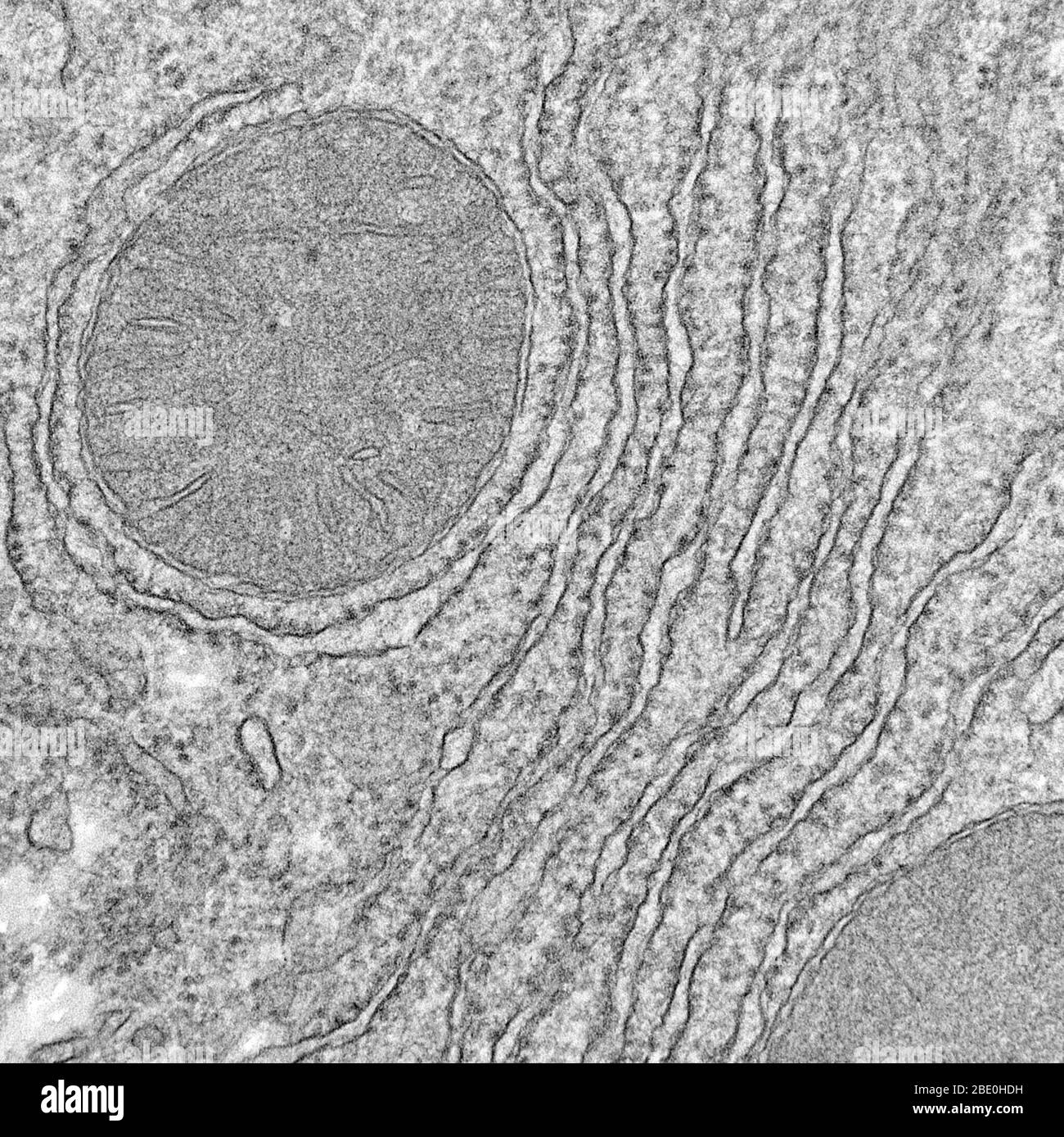
micrograph membrane electron mitochondria reticulum endoplasmic transmission mitochondrion cells microscope organelle eukaryotic visible
Cell Membrane Electron Micrograph
 ar.inspiredpencil.com
Electron Microscope Images Of Cell Membrane
 ar.inspiredpencil.com
Cell Wall Electron Microscope
 mungfali.com
Structure Of Biological Membranes - Functional Organization Of The Cell
 doctorlib.info
cell membrane medical physiology electron micrograph structure 3rd edition transmission biological
Cell Membrane #2 Photograph By Dennis Kunkel Microscopy/science Photo
 fineartamerica.com
membrane microscopy kunkel dennis
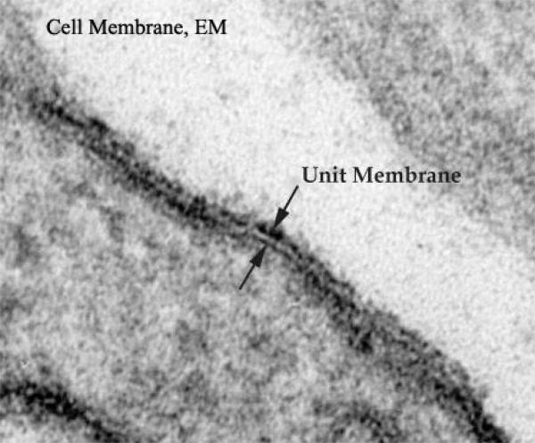
Structure Of The Cell Membrane - Membrane Potential
 www.alpfmedical.info
membrane cell micrograph electron magnification high robertson reproduced acid stained fig courtesy potential structure professor
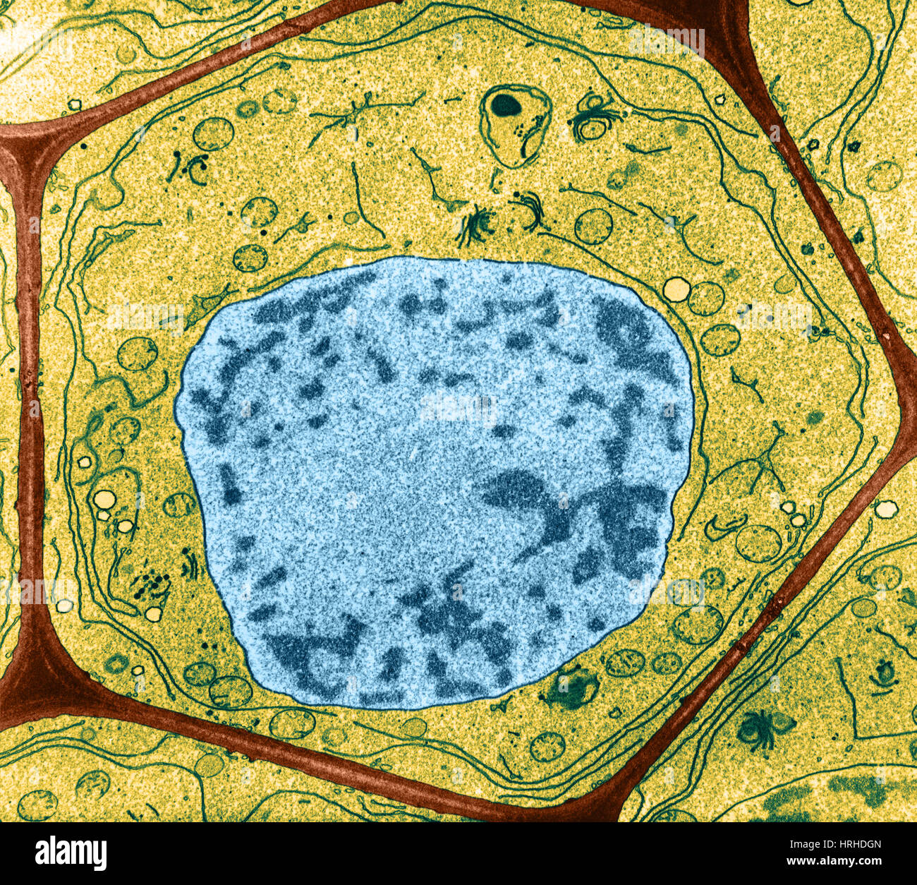
Light Micrograph Of Plant Cells | High-Quality Nature Stock Photos
 creativemarket.com
cells micrograph
Cell Membrane Dr.Jastrow's Electron Microscopic Atlas
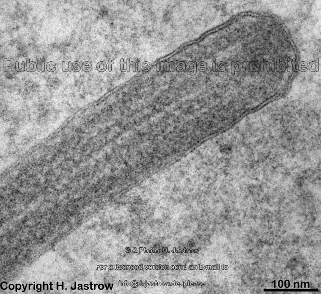 www.drjastrow.de
cell membrane em electron microscopic human
Electron Microscope Images Of Cell Membrane
 ar.inspiredpencil.com
De Fată Egal Hârtie Electron Microscope Images Of Cells Lavandă Se
 notariaurbina.cl
Animal Cell Under Transmission Electron Microscope / Cells Viewed With
 mikegrassere03985.blogspot.com
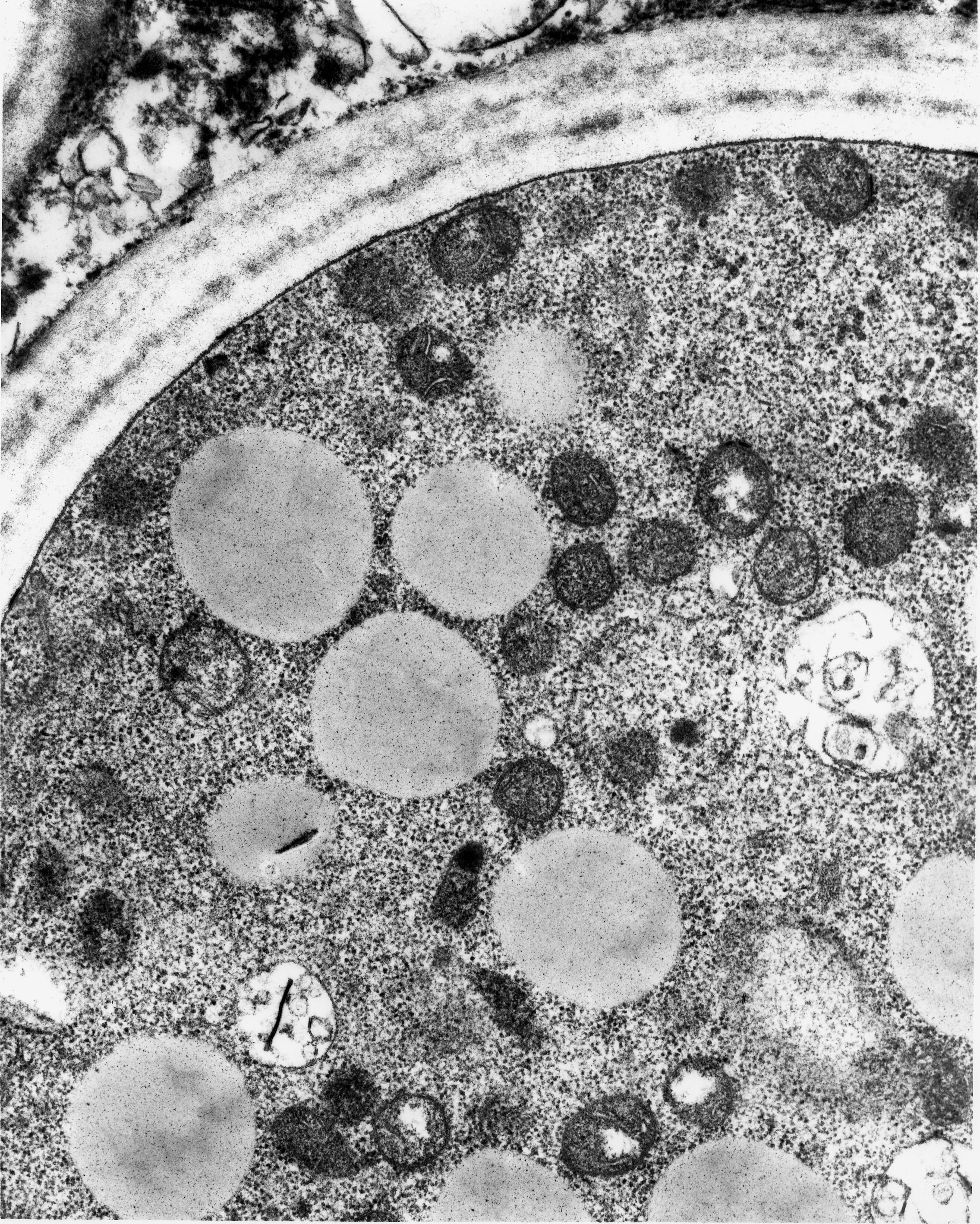
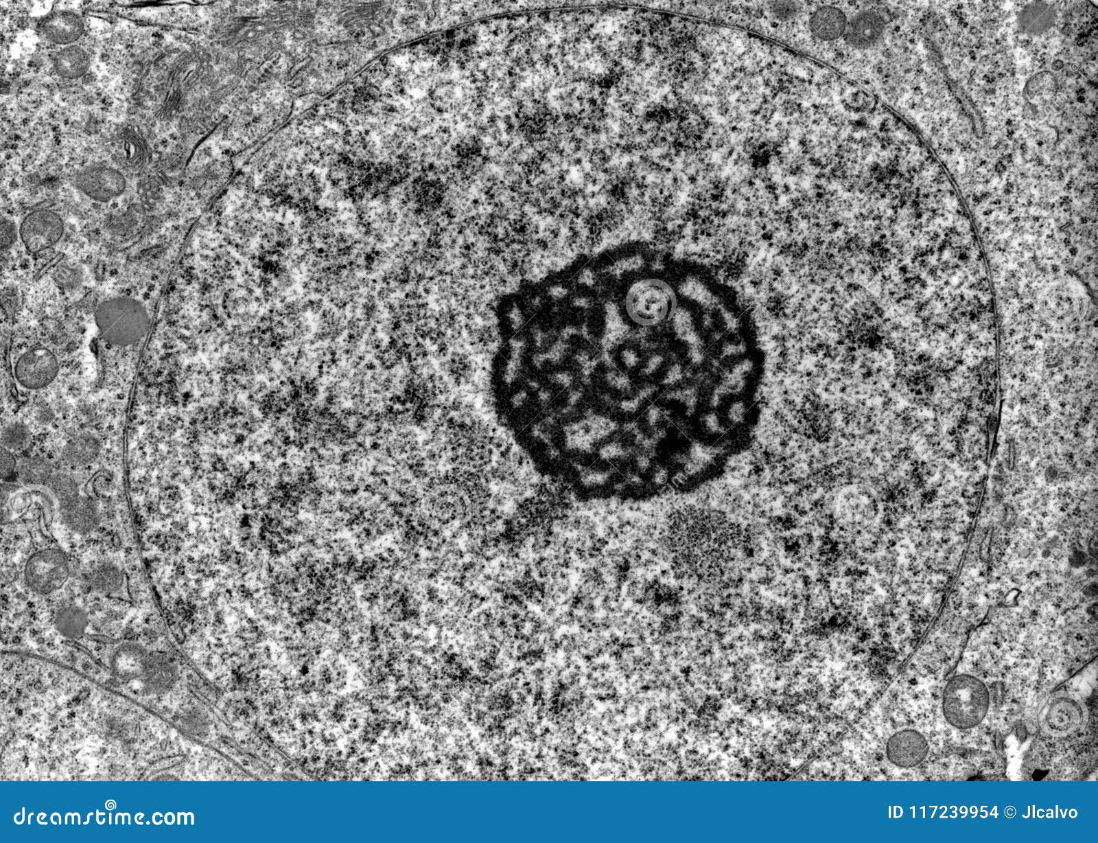
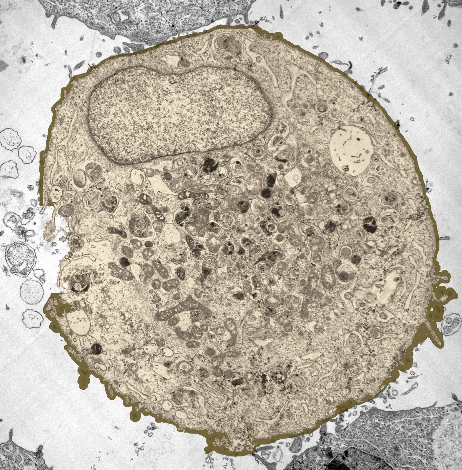
7. Overview Of The Cell | Pocket Dentistry
 pocketdentistry.com
cell electron micrograph membrane nucleus figure overview its visible contents such most pocketdentistry
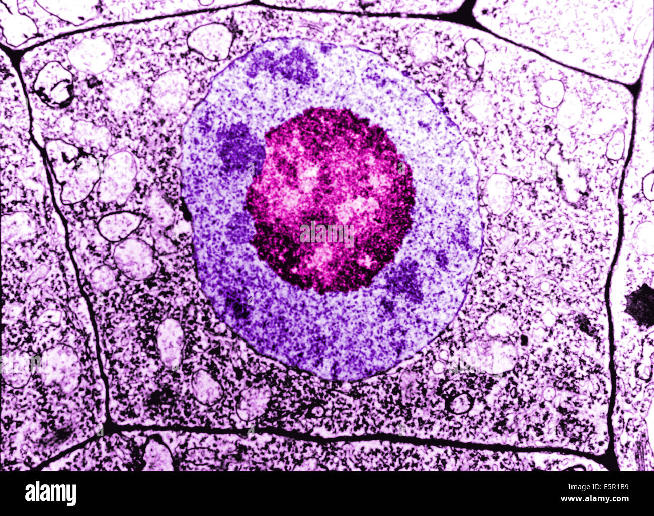
Untitled On Emaze
 app.emaze.com
cell autophagy membrane ovary nuclear cytoplasm mammals like emaze death role intechopen
Cell Micrographs
 science.halleyhosting.com
cells cell prokaryotic micrograph micrographs structure sci chloroplast soph gif description viewed above scale halleyhosting science index
Cell Membrane Micrograph Stock Photos & Cell Membrane Micrograph Stock
 www.alamy.com
cell plant typical omikron alamy stock micrograph wall photograph membrane
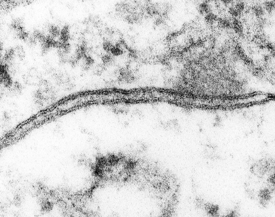
In Clearest View Ever Of Cell Membrane, VCU Team Finds Unexpected
 blogs.vcu.edu
membrane plasmique vcu lipid clearest practitioner bilayer zellmembran mitochondrial finds glance intervju ewert wells seminars darstellung metabolic syndrome processes changes
Membrane Structure - 17 June 2011 - BioInformatics Pakistan
 bioinfopakistan.ucoz.com
membrane cell structure electron cytochemistry biology cilia microscopic function membranes functions flagella celebrate ribosome centrioles knowledge deduce lipoprotein complexes biologists
February 2011 | Cell As A Unit Of Life
 jpsy2011.blogspot.com
cell microscope human under 2011 life february
Elektronenmikroskopie Der Normalen Menschlichen Zellen, Die Zellmembran
 www.alamy.de
electron microscope zellen zellkern membrane microscopy zellmembran cellula nucleo nucleus elektronenmikroskopie normalen menschlichen unterscheiden cellulare normale membrana nucleolus cellule
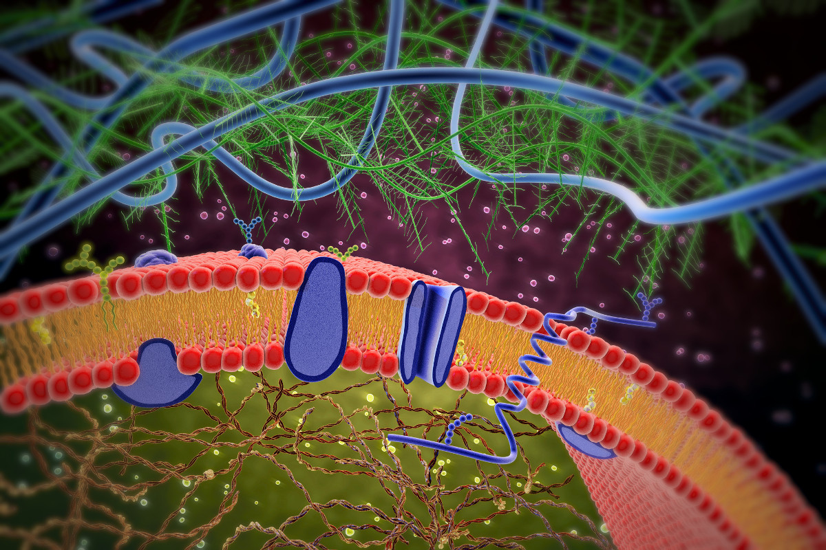
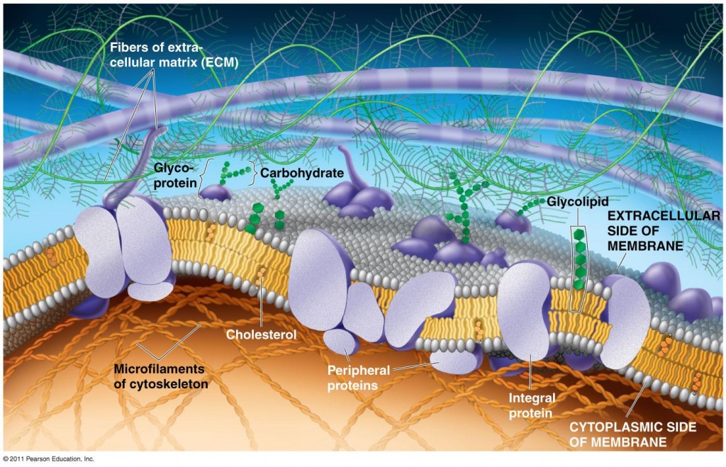
The Plasma Membrane: Structure : Anatomy & Physiology
 anatomyandphysiologyi.com
membrane plasma proteins membrana function biology lipid bilayer fluid functions membranas plasmatica extracellular biologicas fluido mosaico celular funcion bicapa
Cell Membrane Under Electron Microscope - Google Search | Biología
 www.pinterest.com
electron microscopy membrane microscope cell under micrograph plasma animal advanced structure cells bilayer utah plant organelles lipid junction choose board
Cell Membrane Electron Micrograph
 ar.inspiredpencil.com
Membrane Architecture | Celebrate Cytochemistry | Gwen V. Childs, Ph.D.
 www.cytochemistry.net
membrane cell membranes lipid micrograph structure electron plasma microscope bilayer cytochemistry separate showing world phospholipid picture animal archaea bacteria architecture
Label Electron Micrograph Plant Cells
 mavink.com
Electron Microscope Images Of Cell Membrane
 ar.inspiredpencil.com
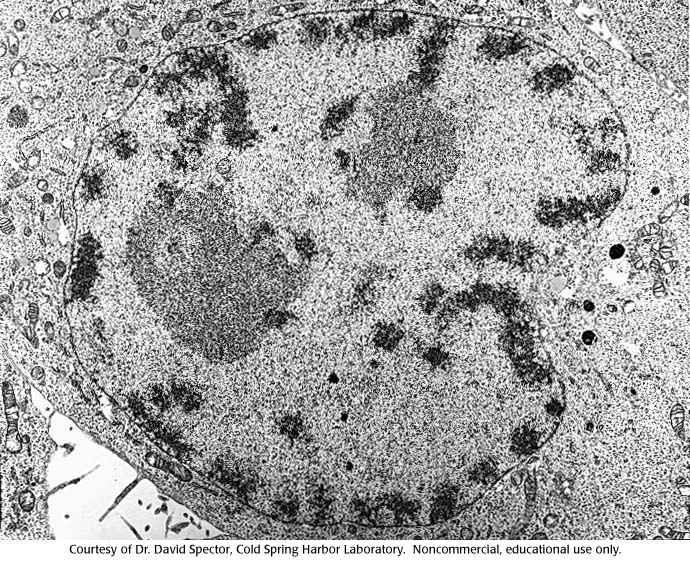
Micrograph Of Cell Dividing, 1 :: CSHL DNA Learning Center
 dnalc.cshl.edu
micrograph dividing
BIOL 230 Lecture Guide - Electron Micrograph Of A Cytoplasmic Membrane
 cwoer.ccbcmd.edu
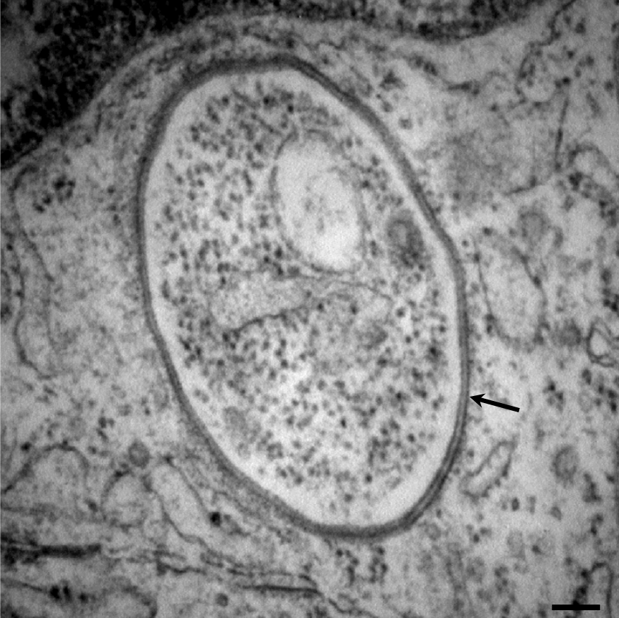
Solved In The Electron Micrograph, Identify The Bacterial | Chegg.com
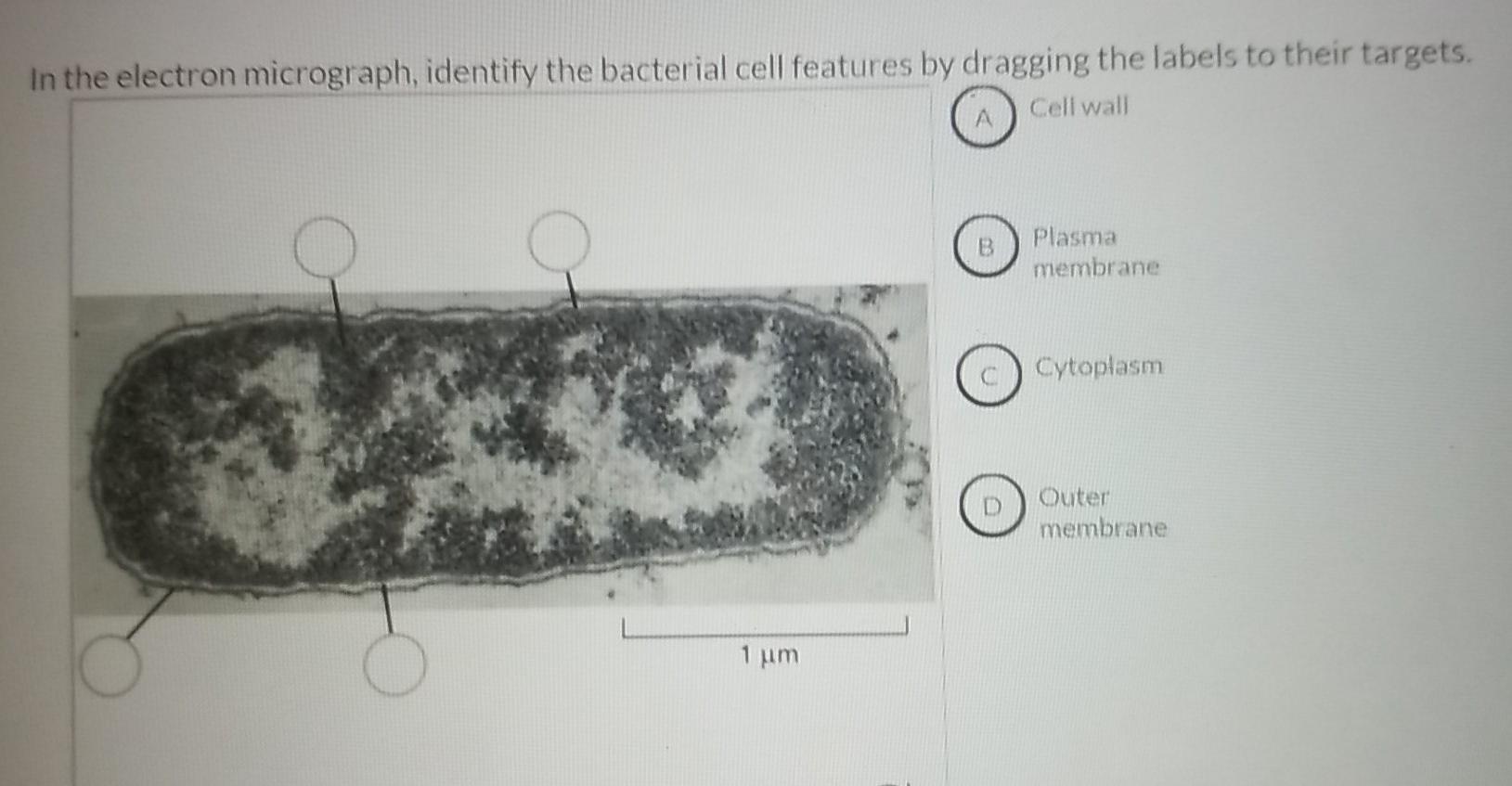 www.chegg.com
De Histology: Phase-Contrast Microscopy & Differential Interference
 dehistology.blogspot.com
cell histology microscopy newest contents comments
The Molecular Biology Of Plant Cells "d0e1050"
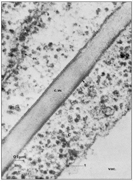 publishing.cdlib.org
cell membrane wall structure cells figure plasmalemma plant membranes publishing ucpressebooks cdlib size full
Electron Micrograph Of Mesangial Cell Plasma Membrane-enriched Fraction
 www.researchgate.net
electron membrane micrograph plasma mesangial fraction enriched microsomes
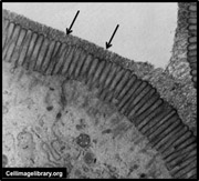
The Cell: The Histology Guide
 histology.leeds.ac.uk
plasma cell membrane histology electron membranes picture cells micrograph dark two intercellular them space shows
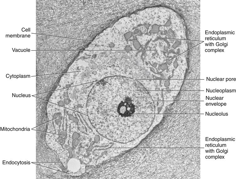
Edexcel IAL Biology: 2.3.3 Describe The Ultrastructure Of An Animal
 ialbio.blogspot.com
cell animal electron organelles structure cells microscope eukaryotic plant under micrograph biology real picture nucleus google diagram nucleolus ribosomes mitochondria
Micrograph of cell dividing, 1 :: cshl dna learning center. Cell membrane em electron microscopic human. Elektronenmikroskopie der normalen menschlichen zellen, die zellmembran